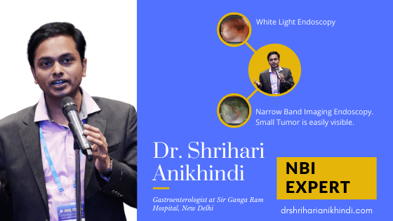
One of the most impressive innovations in the medical field in recent years has been Narrow Band Imaging, also commonly known as NBI. NBI is a modification on conventional endoscopy which is especially useful in detecting subtle findings. It has particularly revolutionized the management of gastrointestinal cancers. To know more about Narrow Band Imaging(NBI) and how it can help you in appropriate diagnosis and therapy, keep reading the whole article.
Birth of an Endoscope
An Endoscope is a medical instrument that can visualize and treat several conditions inside body cavities like the gastrointestinal tract (esophagus, stomach and intestines). The initial endoscopes developed in the 1800s were rigid and very cumbersome to use. In 1932, the first flexible endoscope was developed by Dr.Rudolf Schindler. Being flexible these endoscopes could be easily negotiated inside the body and were hence more acceptable to both doctors and patients.
The last few decades have seen a dramatic improvement in the utility, capability and finesse of endoscope technology. Presently most common endoscope used worldwide is based on the principle of a built-in video camera. The video camera uses a CCD (charged couple device) or HDTV(high definition television) to convert visual images captured from the gastrointestinal tract into electrical signals for display on TV monitor.
White Light Imaging Endoscopy
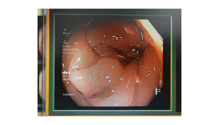
These scopes use white light for visualization of the inner lining of the gastrointestinal tract. An endoscopy done using white light as a source, the image is most commonly seen like this. The mucosa (inner lining of the gastrointestinal tract) appears brownish-yellow on white light endoscopy.
Narrow Band Imaging(NBI) Endoscopy
Recently, exciting innovation in endoscopy technology was invented in Japan. This technology is called image enhanced technology and is based on the principle of enhancing the image by modifying the characteristics of the light source used in the endoscope.
The pioneers in the development and incorporation of this technology were Olympus Corporation Ltd. Based in Tokyo, Japan and they named and patented this technology as Narrow Band Imaging (NBI) in their endoscopes. Subsequently, other medical equipment companies like Fujifilm and Pentax also developed their own image enhancement methods in their endoscopes.
To understand how NBI works, let us understand some principles of light
Principles of Light:
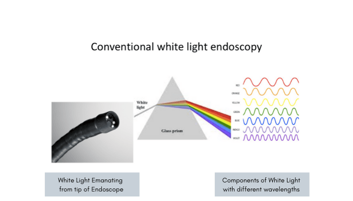
White light is normally made up of a spectrum of 7 colors having different wavelengths (Violet, Indigo, Blue, Green, Yellow, Orange and Red). Suppose a white light is passed through a prism, we can clearly see the components of white light.
Now these individual component colors have different abilities of penetration and refraction depending on their wavelengths. In NBI a special filter is used in the endoscope to allow only the blue and green component of light to pass through.
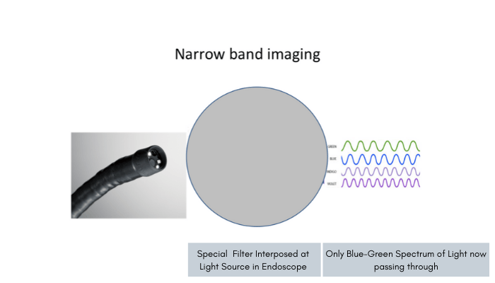
The special property of blue and green light is that they have only superficial penetrance and can enhance the blood vessels on the surface of the mucosa very clearly.
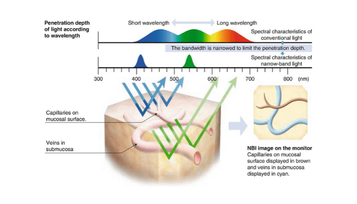
Whenever there is the development of inflammation (like gastritis, H.Pylori infection, Ulcerative Colitis, Crohn’s disease, etc.) and cancer (esophageal cancer, gastric cancer, colonic polyps, colon cancer, etc.) , the first event that occurs is increase in the blood supply to the affected areas. Thus, the concentration of blood vessels increases in the affected mucosa.
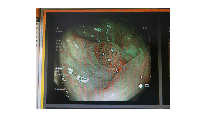
However, these blood vessels are very small and not usually visible to the naked eye using white light endoscopy. However, when the affected area is seen using NBI endoscopy they can be much more clearly identified and delineated as compared to white light endoscopy.
Advantages of Narrow Band Imaging(NBI):
The biggest advantage of using NBI endoscopy is that the affected area can be diagnosed at a very early stage and hence treated easily and completely. It is a well-known fact that if cancer is not detected at an early stage, it is difficult and often impossible to treat.
Several scientific studies done all over the world have shown that NBI is much more effective than a conventional white-light endoscopy in detecting early cancer and various inflammatory conditions. It helps in targeting the high-risk areas for diagnosis and thus enables timely treatment and cure.
Especially for people with risk factors for gastrointestinal cancer like elderly (> 50 years of age), history of smoking, habitual alcohol intake or tobacco chewing, family history of cancer, history of Inflammatory Bowel Disease (Ulcerative Colitis and Crohn’s disease) etc. getting an NBI endoscopy is always recommended over a conventional endoscopy. The biggest advantage is that an NBI endoscopy comes at no significant added cost as compared to conventional endoscopy. The endoscope and the procedure is also similar, only the expertise of the operator matters!
For any queries or further information, please visit the contact us page or send us a mail at contact@drshriharianikhindi.com or keep watching this space.
Or you can subscribe to my YouTube Channel to know NBI and gastrointestinal tract.